Water Quality
Nanotechnology for a Safe and Sustainable use of Water Resources
We work on overcoming some of the most challenging issues in the water environment making responsible use of the great opportunities that nanotechnology offers.
Research Lines:
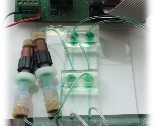
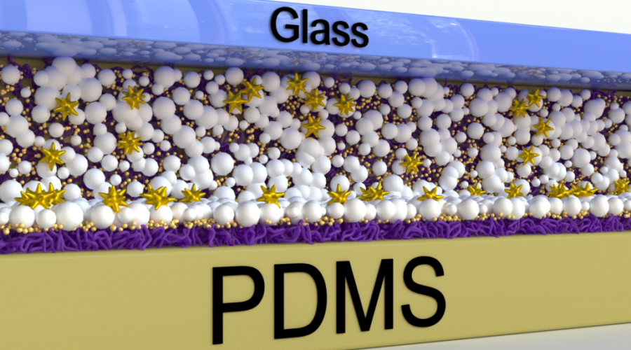
- Nanotech-based sensors for water quality monitoring: we fully develop portable and/or remote biosensors for water biological and chemical contaminants.
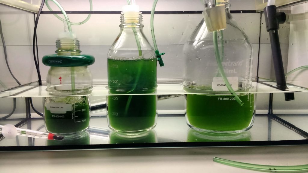
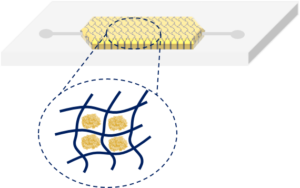
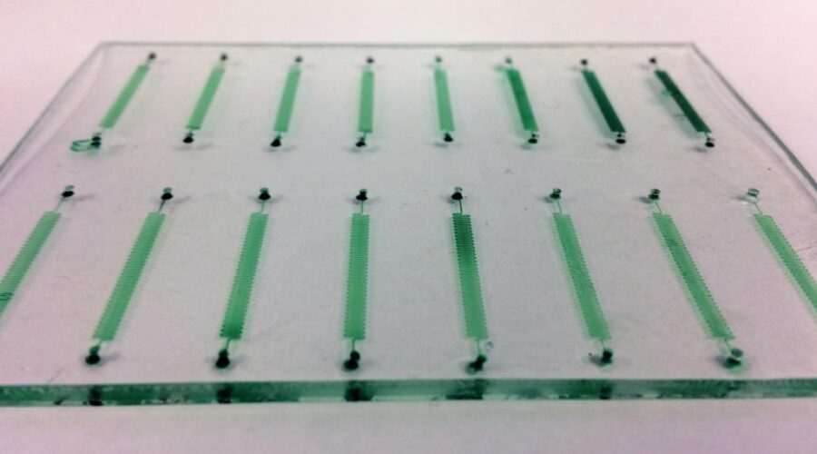
- Nanotechnology for cleaner water: we design, fabricate and test nanomaterials for the selective capture of water contaminants and biotoxins; and micro/nanostructured surfaces to control bacterial adhesion and biofilm formation.
- Environmental risk assessment of nanomaterials: we develop and carry out tests to evaluate nanomaterials’ modification, fate, bioaccumulation and ecotoxicity. We make special emphasis on implementing the safety-by-design concept to the in house produced nanomaterials and from collaborators.


Projects
Publications
-
Evaluation of the Antimicrobial Activity of Chitosan Nanoparticles against Listeria monocytogenes
POLYMERS, 2023One-step synthesis of magnetic covalent organic framework composite for the adsorption of marine toxin okadaic acid
CRYSTENGCOMM, 2023Stable Graphene Membranes for Selective Ion Transport and Emerging Contaminants Removal in Water
ADVANCED FUNCTIONAL MATERIALS, 2023Acute Aquatic Toxicity to Zebrafish and Bioaccumulation in Marine Mussels of Antimony Tin Oxide Nanoparticles
NANOMATERIALS, 2023Assessing Acinetobacter baumannii virulence and treatment with a bacteriophage using zebrafish embryos
FASEB JOURNAL, 2023Single-cell ICP-MS for studying the association of inorganic nanoparticles with cell lines derived from aquaculture species
ANALYTICAL AND BIOANALYTICAL CHEMISTRY, 2023A carboxyl-functionalized covalent organic polymer for the efficient adsorption of saxitoxin
JOURNAL OF HAZARDOUS MATERIALS, 2023A Near-Infrared Mechanically Switchable Elastomeric Film as a Dynamic Cell Culture Substrate
BIOMEDICINES, 2023A novel label-free electrochemical immunosensor for detection of surfactant protein B in amniotic fluid
TALANTA, 2023Cobalt Impregnation on Titania Photocatalysts Enhances Vis Phenol Photodegradation
MATERIALS, 2023Correlative Light, Electron Microscopy and Raman Spectroscopy Workflow To Detect and Observe Microplastic Interactions with Whole Jellyfish
ENVIRONMENTAL SCIENCE & TECHNOLOGY, 2023Getting fat and stressed: Effects of dietary intake of titanium dioxide nanoparticles in the liver of turbot Scophthalmus maximus
JOURNAL OF HAZARDOUS MATERIALS, 2023Green-Based Modifications of Polyethylene Surfaces Enhancing Antifouling Properties in Water-Related Systems
ADVANCED MATERIALS TECHNOLOGIES, 2023Influence of Brewing Process on the Profile of Biogenic Amines in Craft Beers
SENSORS, 2023Innovative Antibacterial, Photocatalytic, Titanium Dioxide Microstructured Surfaces Based on Bacterial Adhesion Enhancement
ACS APPLIED BIO MATERIALS, 2023Layer-by-Layer siRNA Particle Assemblies for Localized Delivery of siRNA to Epithelial Cells through Surface-Mediated Particle Uptake
ACS APPLIED BIO MATERIALS, 2023Submicron- and nanoplastic detection at low micro- to nanogram concentrations using gold nanostar-based surface-enhanced Raman scattering (SERS) substrates
ENVIRONMENTAL SCIENCE-NANO, 2023Bioaccumulation of titanium dioxide nanoparticles in green (Ulva sp.) and red (Palmaria palmata) seaweed
MICROCHIMICA ACTA, 2023Electrochemical determination of heavy metal ions applying screen-printed electrodes based sensors. A review on water and environmental samples analysis
TALANTA OPEN, 2023The Functions of Cholera Toxin Subunit B as a Modulator of Silica Nanoparticle Endocytosis
TOXINS, 2023 -
A novel portable label-free electrochemical immunosensor for ultrasensitive detection of Aeromonas salmonicida in aquaculture seawater
ANALYTICAL AND BIOANALYTICAL CHEMISTRY, 2022Threefold reactivity of a COF-embedded rhenium catalyst: reductive etherification, oxidative esterification or transfer hydrogenation
CHEMICAL COMMUNICATIONS, 2022Towards on-site detection of gluten-containing cereals with a portable and miniaturized prototype combining isothermal DNA amplification and naked eye detection
MICROCHEMICAL JOURNAL, 2022Development of an optical sensor for the determination of perchlorate ions
2022 IEEE International Conference on Design & Test of Integrated Micro & Nano-Systems (DTS), 2022Chapter 1. Fundamentals of Biosensors and Detection Methods
Advances in Experimental Medicine and Biology, 2022Impurities in polyvinylpyrrolidone: the key factor in the synthesis of gold nanostars
Nanoscale Advances, 2022Pitfalls in methods to study colocalization of nanoparticles in mouse macrophage lysosomes
JOURNAL OF NANOBIOTECHNOLOGY, 2022Substrate stiffness reduces particle uptake by epithelial cells and macrophages in a size-dependent manner through mechanoregulation
NANOSCALE, 2022The Choice of Nanoparticle Surface-Coupled Fluorescent Dyes Impacts Cellular Interaction
CHEMNANOMAT, 2022The micro-, submicron-, and nanoplastic hunt: A review of detection methods for plastic particles
CHEMOSPHERE, 2022Toxicity of oleate-based amino protic ionic liquids towards Escherichia coli, Danio rerio embryos and human skin cells
JOURNAL OF HAZARDOUS MATERIALS, 2022Cellular Uptake of Silica and Gold Nanoparticles Induces Early Activation of Nuclear Receptor NR4A1
NANOMATERIALS, 2022Combination of Recombinase Polymerase Amplification with SYBR Green I for naked-eye, same-day detection of Escherichia coli O157:H7 in ground meat
FOOD CONTROL, 2022Proteomics reveals multiple effects of titanium dioxide and silver nanoparticles in the metabolism of turbot, Scophthalmus maximus
CHEMOSPHERE, 2022Label-free SERS techniques in biomedical applications
SERS for Point-of-care and Clinical Applications (ELSEVIER), 2022pH-sensitive nanoliposomes for passive and CXCR-4-mediated marine yessotoxin delivery for cancer therapy
NANOMEDICINE, 2022 -
Study on the efficiency of a covalent organic framework as adsorbent for the screening of pharmaceuticals in estuary waters
CHEMOSPHERE, 2021Additional Commentary on the Detection and Quantification of Plastic Micro- and Nanoparticles in Tea Samples
CHIMIA, 2021Plastics in our ocean as transdisciplinary challenge
MARINE POLLUTION BULLETIN, 2021Detection of Silver Nanoparticles in Seawater Using Surface-Enhanced Raman Scattering
NANOMATERIALS, 2021Acute ecotoxicity assessment of a covalent organic framework
ENVIRONMENTAL SCIENCE-NANO, 2021Aligned and Oriented Collagen Nanocomposite Fibers as Substrates to Activate Fibroblasts
ACS APPLIED BIO MATERIALS, 2021Combining microcavity size selection with Raman microscopy for the characterization of Nanoplastics in complex matrices
SCIENTIFIC REPORTS, 2021Covalent organic framework as adsorbent for ultrasound-assisted dispersive (micro)solid phase extraction of polycyclic synthetic fragrances from seawater followed by fluorescent determination
ANALYTICA CHIMICA ACTA, 2021Selection of Covalent Organic Framework Pore Functionalities for Differential Adsorption of Microcystin Toxin Analogues
ACS APPLIED MATERIALS & INTERFACES, 2021 -
A SERS-based 3D nanobiosensor: towards cell metabolite monitoring
MATERIALS ADVANCES, 2020Covalent Organic Framework Composites: Synthesis and Analytical Applications
MOLECULES, 2020Efficient adsorption of endocrine-disrupting pesticides from water with a reusable magnetic covalent organic framework
MICROPOROUS AND MESOPOROUS MATERIALS, 2020Evaluation and implementation of commercial antibodies for improved nanoparticle-based immunomagnetic separation and real-time PCR for faster detection of Listeria monocytogenes
JOURNAL OF FOOD SCIENCE AND TECHNOLOGY-MYSORE, 2020Improved Photocatalyzed Degradation of Phenol, as a Model Pollutant, over Metal-Impregnated Nanosized TiO2
NANOMATERIALS, 2020Improved Photocatalyzed Degradation of Phenol, as a Model Pollutant, over Metal-Impregnated Nanosized TiO2 (vol 10, 996, 2020)
NANOMATERIALS, 2020Extraction of Ibuprofen from Natural Waters Using a Covalent Organic Framework
MOLECULES, 2020Particle Surfaces to Study Macrophage Adherence, Migration, and Clearance
Advanced Functional Materials, 2020A comparative study of silver nanoparticle dissolution under physiological conditions
NANOSCALE ADVANCES, 2020Are TiO2 nanoparticles safe for photocatalysis in aqueous media?
NANOSCALE ADVANCES, 2020Chapter 2. New techniques in environment monitoring.
Climate Change and Marine and Freshwater Toxins. De Gruyter, 2020Magnetic Hyperthermia for Cancer Treatment: Main Parameters Affecting the Outcome of In Vitro and In Vivo Studies
MOLECULES, 2020Multifuntional Gold Nanoparticles for the SERS Detection of Pathogens Combined with a LAMP-in-Microdroplets Approach
MATERIALS, 2020 -
Gold Nanostars for the Detection of Foodborne Pathogens via Surface-Enhanced Raman Scattering Combined with Microfluidics
ACS APPLIED NANO MATERIALS, 2019Recyclable magnetic covalent organic framework for the extraction of marine biotoxins
NANOSCALE, 2019Electrochemical Immunosensor for TNF alpha-Mediated Inflammatory Disease Screening
ACS CHEMICAL NEUROSCIENCE, 2019Polymer-Coated Gold Nanospheres Do Not Impair the Innate Immune Function of Human B Lymphocytes in Vitro
ACS NANO, 2019Phase Transformation of Superparamagnetic Iron Oxide Nanoparticles via Thermal Annealing: Implications for Hyperthermia Applications
ACS APPLIED NANO MATERIALS, 2019A hydrofluoric acid-free method to dissolve and quantify silica nanoparticles in aqueous and solid matrices
SCIENTIFIC REPORTS, 2019BSA/ASN/Pol407 nanoparticles for acute lymphoblastic leukemia treatment
BIOCHEMICAL ENGINEERING JOURNAL, 2019Effectiveness and Safety of a Nontargeted Boost for a CXCR4-Targeted Magnetic Hyperthermia Treatment of Cancer Cells
ACS OMEGA, 2019Microporous Plasmonic Capsules as Stable Molecular Sieves for Direct SERS Quantification of Small Pollutants in Natural Waters
CHEMNANOMAT, 2019Nanoparticle administration method in cell culture alters particle-cell interaction
SCIENTIFIC REPORTS, 2019Nanoparticle Behaviour in Complex Media: Methods for Characterizing Physicochemical Properties, Evaluating Protein Corona Formation, and Implications for Biological Studies
978-3-030-12461-8, 2019Portable sensing system based on electrochemical impedance spectroscopy for the simultaneous quantification of free and total microcystin-LR in freshwaters
BIOSENSORS & BIOELECTRONICS, 2019Tailoring Covalent Organic Frameworks To Capture Water Contaminants
CHEMISTRY-A EUROPEAN JOURNAL, 2019A Bio-Inspired Amplification Cascade for the Detection of Rare Cancer Cells
CHIMIA, 2019Artificial Lysosomal Platform to Study Nanoparticle Long-term Stability
CHIMIA, 2019Biocompatibility and bioimaging potential of fruit-based carbon dots
NANOMATERIALS, 2019Quantification of Carbon Nanotube Doses in Adherent Cell Culture Assays Using UV-VIS-NIR Spectroscopy
NANOMATERIALS, 2019