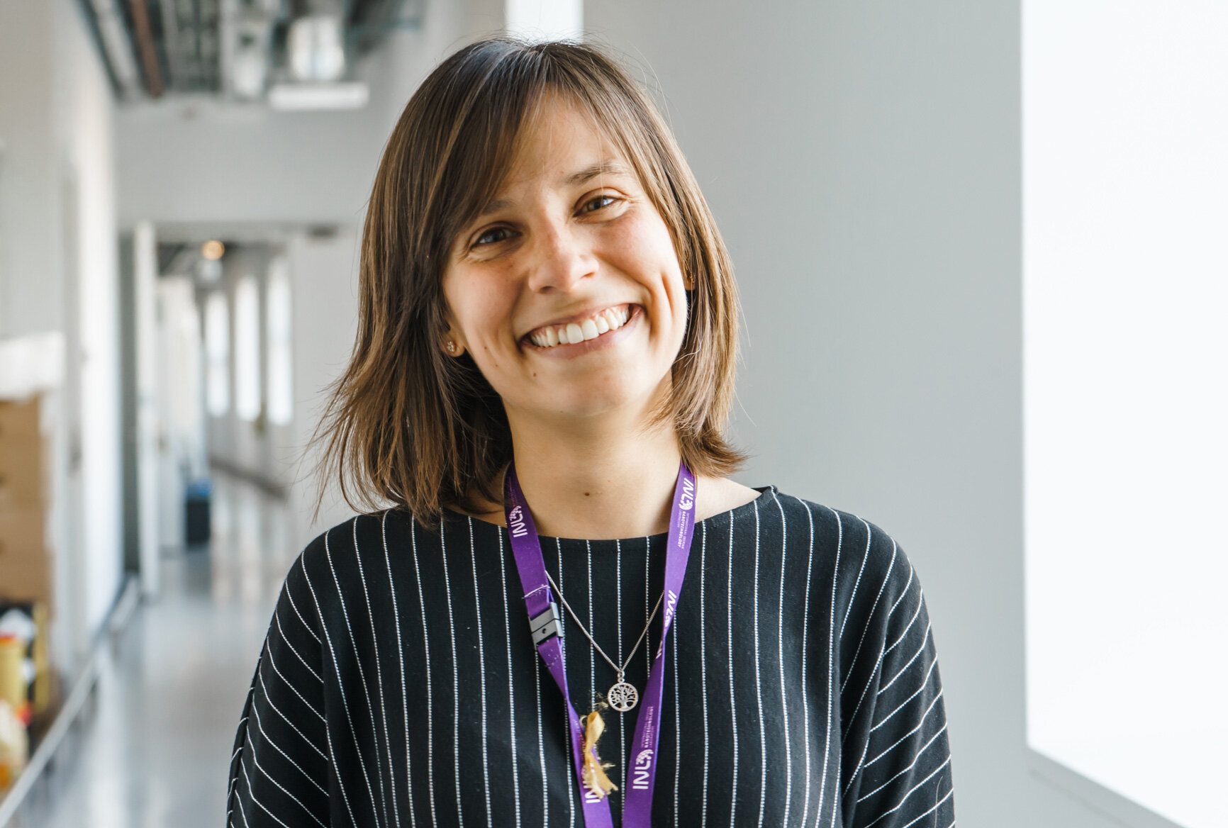
Celebrating WOMEN IN SCIENCE: Interview with Catarina Moura
February 8, 2020
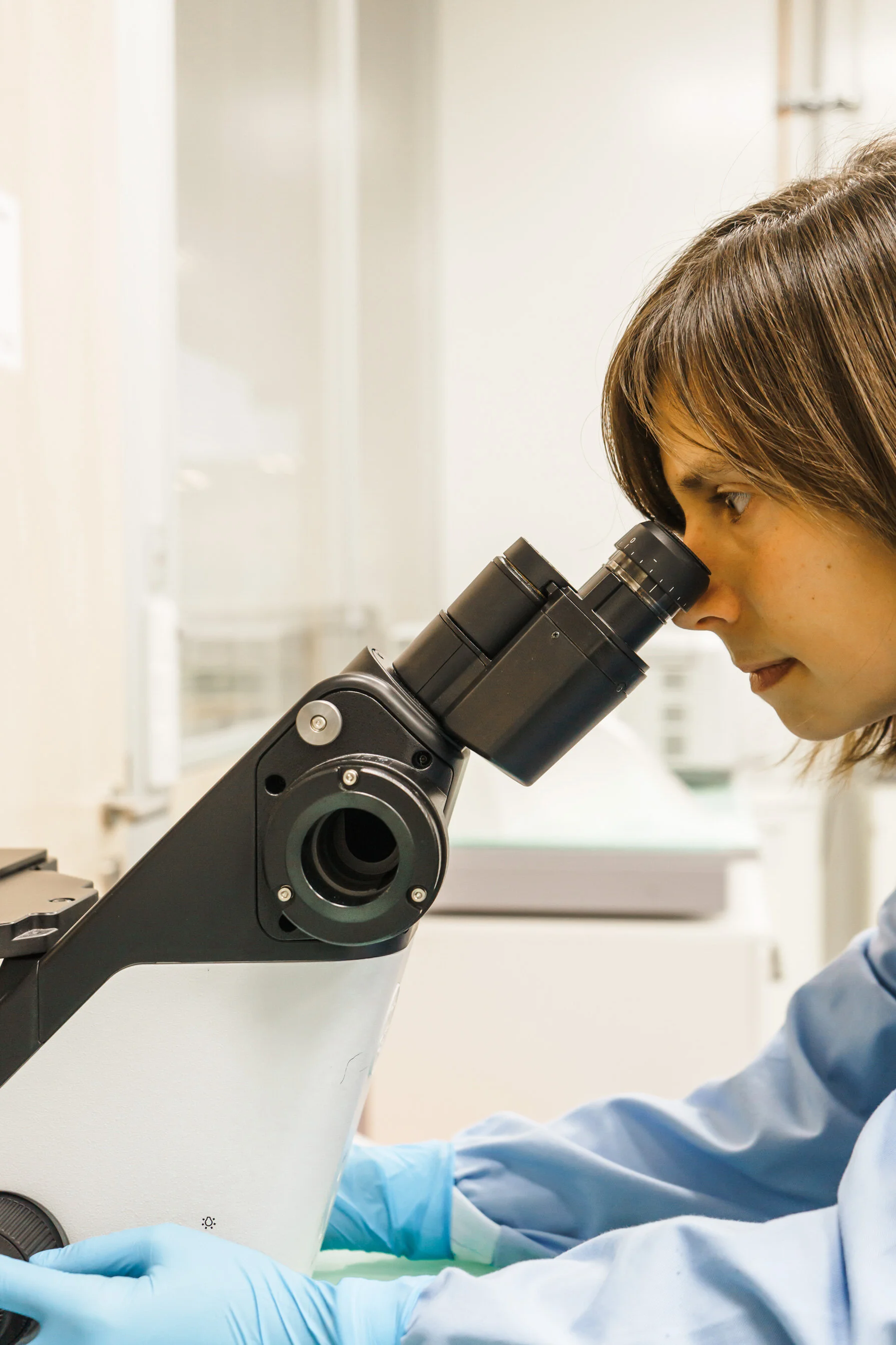
Catarina Moura is a Research Fellow from the INL Medical Devices group within the Department of Life Sciences. She is working on a combined microscopy platform integrating Structured Illumination Microscopy (SIM) and Atomic Force Microscopy (AFM) and applying it to answer outstanding questions regarding cellular signalling.
Catarina is interested in making science accessible and fun to everyone, and she has also been involved in several science outreaches and public engagement activities.
What got you interested in the Life Sciences – Medical Devices?
I’ve always liked to explore – being outdoors, travelling, cooking with new ingredients, learning about new cultures… I believe my curiosity about life sciences combined with my hands-on way of doing things pushed me to study bioengineering. I gained an extensive and multi-disciplinary education, covering several subjects including biology, chemistry, materials science, and tissue engineering, which has undoubtedly increased my interest and enthusiasm in the study of new biomedical approaches.
What is the importance of your research?
Throughout the last century, medical breakthroughs have led to a great increase in life expectancy. However, as a consequence, an increasingly ageing population has led to an increase in age-related diseases causing a dramatic impact on healthcare.
Microscopes are an essential instrument in the biomedical field – for example, for clinical diagnostics or identification of different types of cells in a tumour. Effective medical approaches are essential to fulfil the current demographic challenges, and multimodal imaging methods are emerging as potential alternatives in biomedicine.
We are currently taking the first steps for combining different microscopy technologies in a single imaging platform, so we can assess complementary information using the same sample.
What is the most exciting discovery you made during your research?
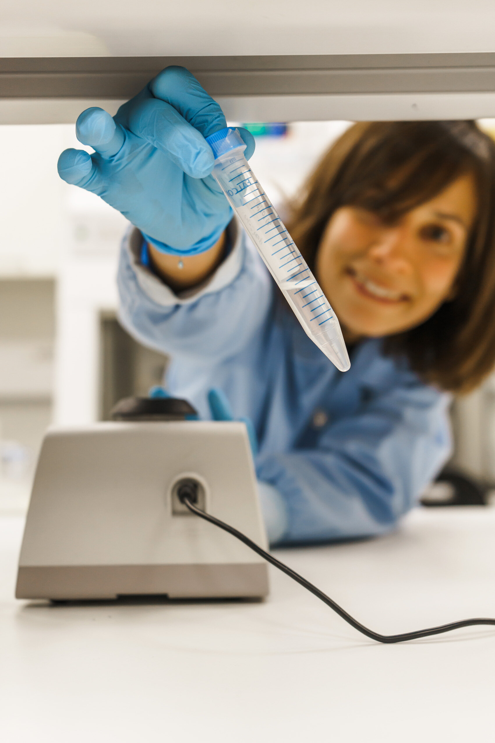
I have been working on a combined microscopy system developed at INL (atomic force microscopy with differential spinning disk fluorescence imaging), and I have demonstrated that this technique allows for the detection of relevant properties of living cells, in real-time.
More specifically, our preliminary work opens the possibility to study the mechanical properties of individual cell populations in samples that cannot be classified and discriminated using morphology alone. This might be important in complex samples where 100% cell purity cannot be achieved or in scenarios where the interplay between different cell types alters the mechanical properties of individual cells (e.g. metastatic cancer cells).
Briefly, what excites you about your work and INL?
One of the things that sparkled my attention to work at INL was interdisciplinarity. I believe interdisciplinary studies foster the creation of new ideas and strategies, not only in the lab but also in everyday work situations. At INL I have also had the opportunity to participate in science communication activities which definitely help release creativity and unlock new collaborations and resources.