Nanophotonics & Bioimaging
The Nanophotonics & Bioimaging (NBI) facility features cutting-edge optical instrumentation and the know-how required for the characterisation of material and life sciences samples.
The INL expert team provides knowledge in the analysis of light-matter interactions at various scales: reaching from the detection of individual fluorescent molecules or nanoparticles to the high-resolution label-free and fluorescence-based imaging of fixed and live cells, e.g. in 2D cell models or tissues.
Furthermore, we are experienced in designing optically active nanomaterials and characterising e.g. plasmonic interaction effects or surface-enhanced Raman effects (SERS) and studying optical properties of metamaterials.
Operating as an open-access facility, we provide support in training and service measurements on spectroscopy and imaging equipment, as well as complementary services, such as specimen/sample preparation, flow cytometry, and spectral or image analysis. Since 2020, the NBI facility has been integrated into the Portuguese Platform of Bioimaging (PPBI).
New generation imaging and spectroscopy solutions that:
- Are characterised by multimodality for faster and more comprehensive analysis of your samples and allowing correlative imaging analysis
- Involve the use of INL-developed innovative contrast agents
- Allow optical super-resolution – surpassing the typical few hundreds of nanometre resolution in diffraction-limited classical microscopy and approaching the nanometre scale.


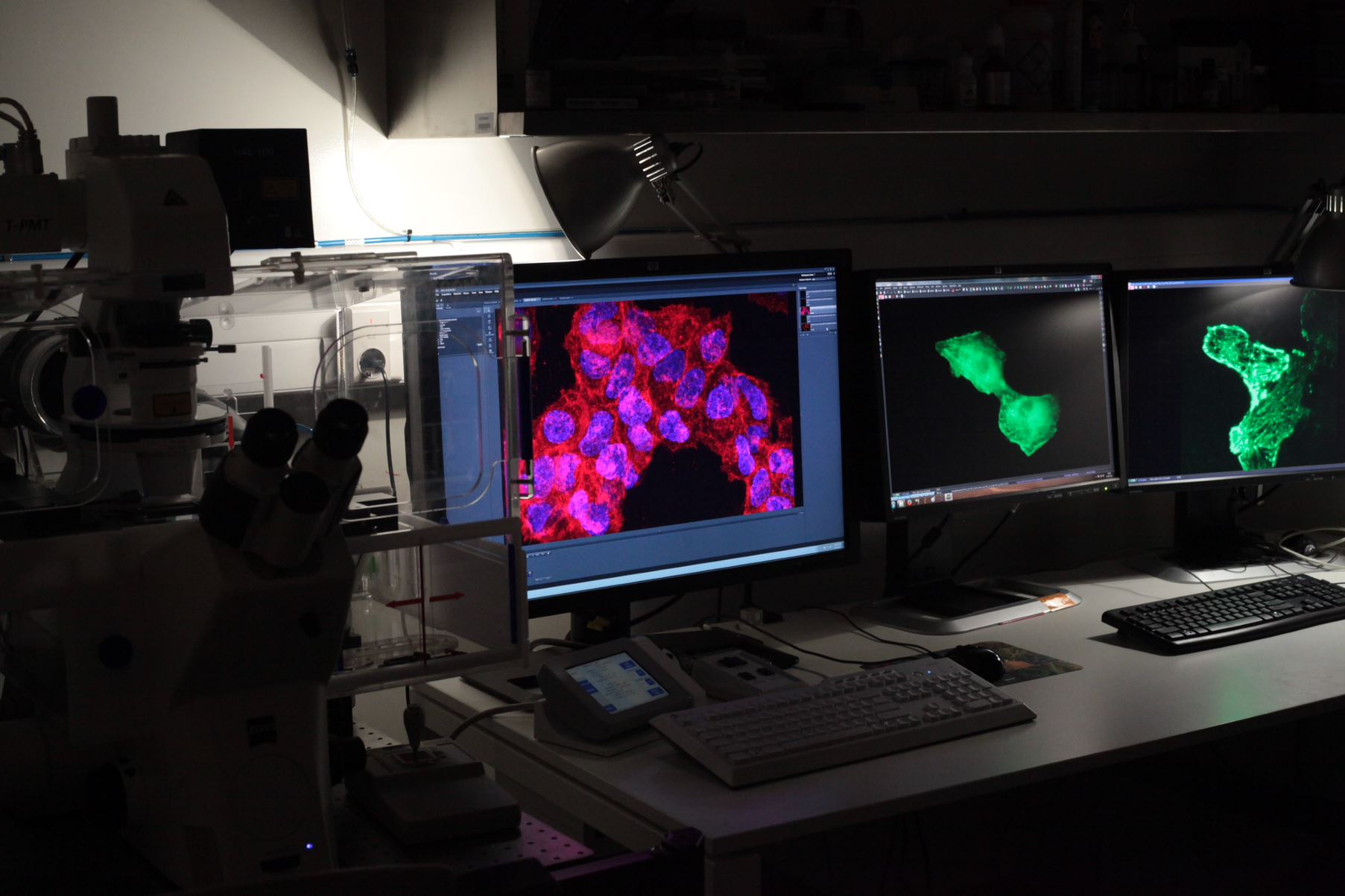
Confocal Fluorescence Microscope (Zeiss, LSM 780) – Used to acquire optical sections with high contrast, high resolution in x, y and z, and allowing high sensitivity in life sciences imaging. Equipped with systems to perform Lambda Scan, and different types of correlation spectroscopy (FCS/FCCS and RICS), as well as the registry of phase contrast, DIC and polarized laser scanning images. This setup is equipped with 6 Laser lines (405, 458, 488, 514, 561, and 633 nm), 5 Zeiss objectives (10x/0.3; 20x/0.8; 40x/1.3 oil; 40x/1.2 water for FCS; 63x/1.40 oil), and an environmental chamber for live imaging.
Holotomography microscope (Nanolive, 3D Cell-Explorer) – Used no acquire label-free 3D images of living cells in physiological conditions (close incubation chamber) without any bleaching or phototoxicity, and without the need of stains or fixation. The generated non-invasive holotomographs, obtained using the phase-shift of light in many cross sections to achieve 3D imaging, show with details the cellular structures and allow recording cellular processes in real time, for long periods (up to weeks).
Fluorescence Microscope (Nikon, Ti-E TIRF) – Used to acquire high-resolution fluorescence widefield images. An inverted total internal reflection fluorescence microscope works by confining excitation energy to a thin section, therefore high signal-to-noise-ratio is obtained. Together with a fast camera (Andor DU-897), it is possible to do dSTORM and observe single molecule fluorescence with this setup. It is fitted with 6 objectives (10x/0.3; 20x/0.5; 40x/0.8; 60x/1.4 oil; 60x/1.5 oil for TIRF; 100x/1.4 oil), 4 Lasers (405, 488, 561, and 633 nm), fluorescence and white light sources, a colour camera (Nikon DS- Fi2) and environmental chamber for live imaging. Dark-field, phase contrast and DIC modes are also available.
Confocal Raman Microscope (Witec, Alpha300 R) – Combining vibrational/Raman spectroscopy with microscopy, this setup is used to obtain hyperspectral image with the information of the Raman spectrum at every pixel, thus the component’s distribution in a sample can be analysed. Fitted with 3 objectives (10x/0.2; 50x/0.7; 100x/0.9), 3 lasers (532, 633 and 785 nm), 600g/mm and 1800g/mm diffraction gratings, and UHTS300 spectrometers coupled to Andor Peltier cooled CCD detectors, this tool is mostly used for analysis of graphene-based devices and structures, characterizing crystallinity, polymorphism, orientation, mechanical stress, as well as contaminants in 2D samples.
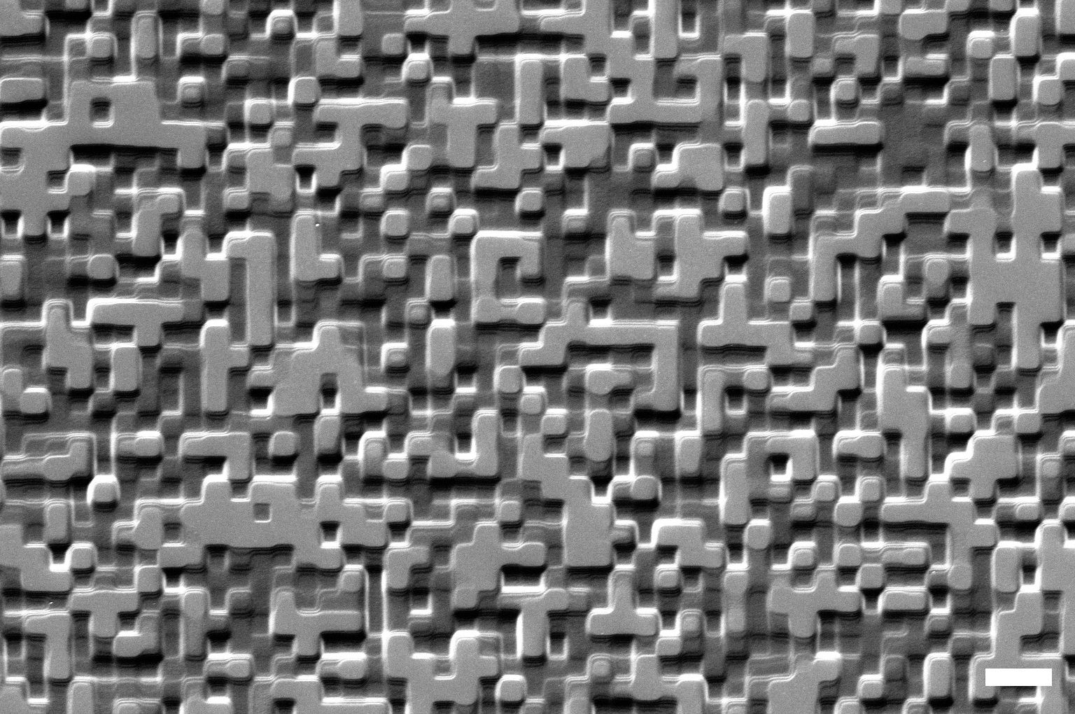
Atomic force Microscopy (Bruker, Dimension Icon AFM) – Used to obtain topographic imaging, this is a scanning probe microscopy that can reach atomic-level resolution. The sharp tip of the mechanical probe (cantilever) makes contact to the sample and is scanned throughout the surface. The bending movements are recorded via the intensity of a laser, reflected from the back of the cantilever into a photodiode. Besides topological information, this setup can make force measurement and manipulate the sample via its interaction with the cantilever tip. Other attributes, such as mechanical (stiffness or adhesion strength) and electrical (conductivity or surface potential) properties, can also be measured locally and displayed within a multidimensional image.
Upright Fluorescence Microscope (Nikon, Ni-E) – Direct epifluorescence microscope for inspection of organic and inorganic materials and devices. Equipped with 6 objectives (1x/0.03; 5x/0.15; 20x/0.45; 50x/0.6; 60x/1.4 oil; 100x/0.8), a Nikon DS- Fi2 and a Hamamatsu Orca-R2 cameras, diascopic and episcopic white light sources, and the registry of phase contrast and polarized images.
Inverted Fluorescence Microscope (Nikon, Ti-E) – Inverted epifluorescence microscope, specially adapted for imaging microfluidics devices. Equipped with an Andor NEO sCMOS Camera, long work-distance objectives (10x/0.30 WD= 16; 20x/0.50 WD=8.2; 60x/0.7 WD=2.6), diascopic and episcopic white light sources, motorized focal adjustment and sample positioner.
Upright Microscope (Nikon, LV100 ND) – Direct microscope for inspection. This microscope is fitted with 5 objectives (5x/0.15; 10x/0.30; 20x/0.45; 100x/0.9), a Nikon DS-Fi2 camera, diascopic and episcopic light sources, allowing to obtain bright field, dark field, phase contrast, DIC and polarized images.
Inverted Microscope (Nikon, MA200) – Inverted microscope with episcopic illumination for inspection. Equipped with 3 objectives (5x/0.15; 10x/0.3; 20x/0.45) and a Nikon DS-Vil camera. This microscope can record bright field, dark field and polarized image. A fluorescence light-source access and filter sets for fluorescence imaging are also available under request. This system was especially adapted to record high velocity videos with an extra high-speed camera and light source.
PerkinElmer, LAMBDA 950 UV-VIS-NIR Spectrophotometer – Used to measure transmission, absorption or reflectance (with accessory) spectra of liquid and solid samples from 175nm to 3300nm. This tool is suited for chemistry and material science applications, measuring liquids, gels, and solid materials important data for sample analysis, and quality control. With different detection accessories (2D – PMT and PbS detectors; 60mm integrating sphere – Spectralon® coated, PMT and InGaAs detectors), this tool has the flexibility and sensitivity to perform a plethora of experiments.
SHIMADZU, UV-2600i Spectrophotometer – This single monochromator system is used to measure transmission or absorption spectra from 185nm to 900nm. This tool is suited for material science applications, measuring important data for sample analysis, and quality control.
Horiba, FluoroMax-4 Compact Spectrofluorometer – High performance tabletop fluorometer to measure excitation and emission spectra, fluorescence and phosphorescence. Allowing the characterization of physicochemical properties of samples, to obtain information about the surrounding environment.
ISS Chronos BH, Fluorescence lifetime, emission, excitation and anisotropy setup – The ISS Chronos BH is a time-domain fluorometer with picosecond resolution. Its optical design and automatic instrument control are state-of-the-art for time-resolved fluorometers.
Jasco J-815 Circular Dichroism Spectroscopy System – Obtain structural information in chiral compounds, such as unfolding and folding, protein-ligand binding and RNA/DNA interaction.
Bruker, Vertex 80v vacuum FTIR Spectrometer – Fourier Transformed Infrared Spectroscopy. Fitted with different accessories, allowing ATR and transmission detection. Used in quality control to verify the identity and specifications of raw materials and products. Identifying compounds or investigating the chemical composition of a sample.
J. A. Woollam, M2000 Spectroscopic Ellipsometer – Accurate and versatile ellipsometer for research on all types of materials: semiconductors, dielectrics, polymers, metals, multi-layers, and more. Measurement of optical properties of thin materials such as transparent layers, thin films and coatings.
Accurion, Nanofilm-ep4 Spectroscopic Imaging Ellipsometer – Used for localized measurements of the optical properties of thin materials such as transparent layers, thin films and coatings, as well as liquid-solid interface.
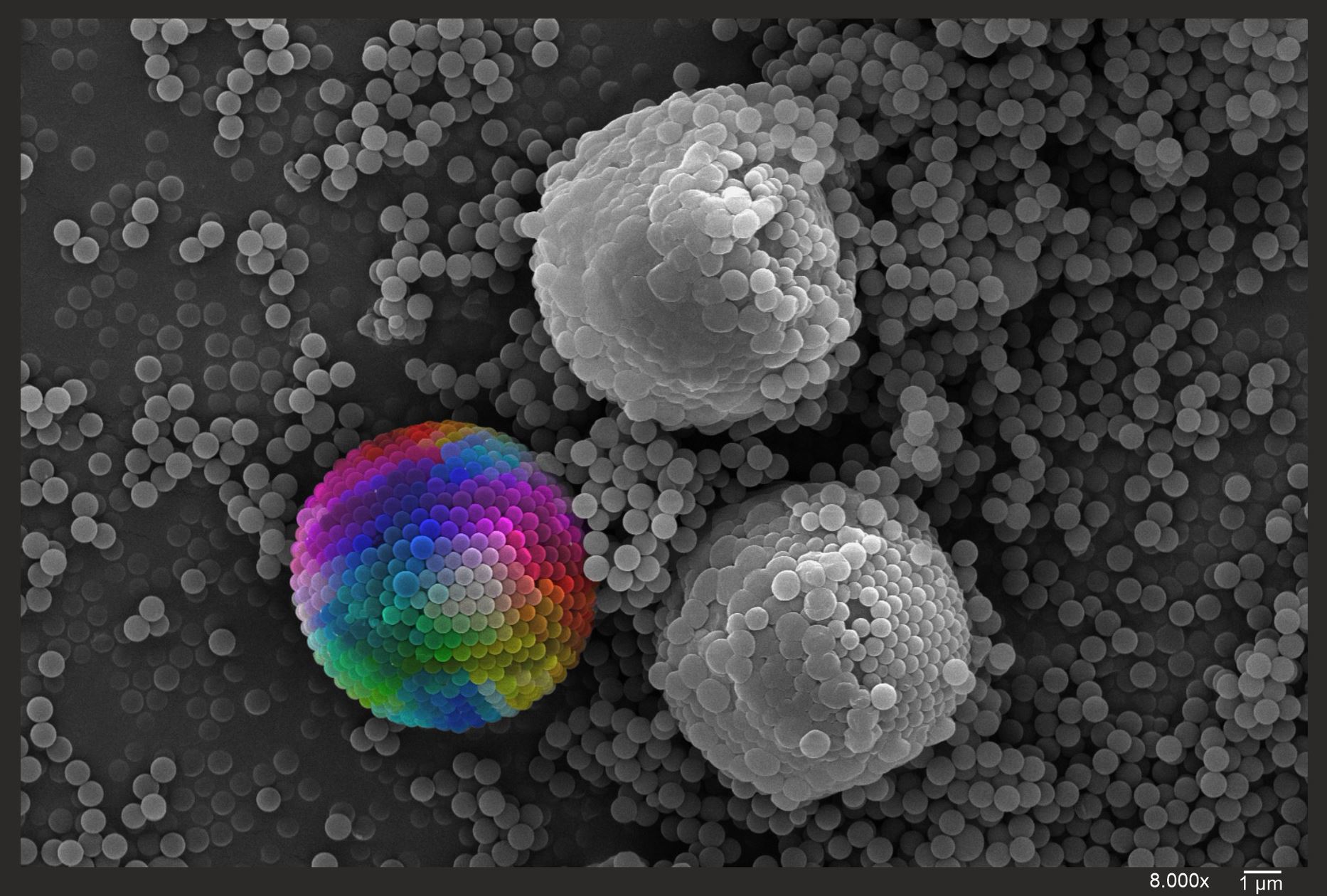
Dynamic Light Scattering – DLS (Horiba, SZ-100Z )– Measure particle size and zeta potential by dynamic light scattering. Characterization of colloidal solutions, providing important information about the surface charge and size distribution of particle and emulsions.
Nanoparticle Tracking Analysis – NTA (Malvern Panalytical, NanoSight NS300) – Imaging Nanoparticle Tracking Analysis. Allowing visualization and measurement of particle size and concentration in liquid suspension.
Gel imaging fluorescence system (Synergy, G:BOX CHEMI XT4) – Gel imaging fluorescence system mostly used to detect proteins on western blots assays. Chemiluminescence is used for sensitive detection of picogram or femtogram amounts of materials, while fluorescence is used to quantify and detect different proteins on one blot. This tool is fitted with different filters for standard fluorescence applications or for chemiluminescence imaging.
Synergy, Biotek H1 Microtiter Plate Reader – Measure chemical, biological or physical reactions, properties and analytes within the wells of a microplate. Providing top and bottom fluorescence intensity, UV-visible absorbance and luminescence detection. Mostly used to quantify biological and chemical assays.
BioRad, S3-e Flow Cytometry and Cell Sorter – Flow cytometry is a sophisticated technique to measure multiple physical characteristics of a single particle such as size and granularity simultaneously as the particles flows in suspension through a measuring device. Its working depends on the light scattering features of the sample. This tool analyses physical and chemical characteristics of isolated cells, biological or non-biological particles. It also counts cells or particles based on physical-chemical characteristics and quantify these cellular properties. Besides characterising the sample, this machine can sort the particles depending on specific characteristics such as size and fluorescence.
Combined AFM and fluorescence microscope (JPK, NanoWizard3 AFM combined with, Fluorescence microscope Nikon, Ti-S/L100)– Designed to provide fluorescent microscopy images and the highest atomic force microscopy performance in liquids and air. Measure and monitor biological samples, correlating biochemical identification, via fluorescence, with surface and mechanical characterization. Fitted with a 60x/1.4 oil objective (other objectives are also available), allows optimum imaging in air and liquid for single molecules, polymers and nanomaterials. (contact: Pieter.deBeule@inl.int, Adelaide.Miranda@inl.int )
Femtosecond laser-based 3D microfabrication (contact: Jana.Nieder@inl.int , Christian.Maibohm@inl.int )
Hamamatsu, Universal Streak Camera system integrated in custom, optical setups for cuvette and / or droplet/ surface measurements – when coupled to an inverted microscope platform. (Contact: Jana.Nieder@inl.int )
Multiphoton, Second Harmonic Generation and FLIM – Custom developed fluorescence lifetime imaging microscope with single molecule sensitivity, suited for life cell observation, fluorescence anisotropy detection and nanomedicine research. Anisotropy and Hanbury Brown Twiss detection schemes implemented as well as possibility to couple the output to a Streak detection system. The system can be coupled with femtosecond and picosecond Lasers, different CCD cameras, fast electronics for Time Correlated Single Photon Counting, filters for one and multiphoton excitation/detection modes, and incubator for live cell experiments. (contact: Jana.Nieder@inl.int , Christian.Maibohm@inl.int )
Metal-induced energy transfer fluorescence lifetime imaging (MIET-FLIM) (Contact: Jana.Nieder@inl.int ). This technique uses custom developed cover slips fabricated at INL clean room facilities.
Intracellular temperature microscopy (Contact: Jana.Nieder@inl.int )
ODMR upgrade for widefield TIRF microscope (Contact: Jana.Nieder@inl.int)