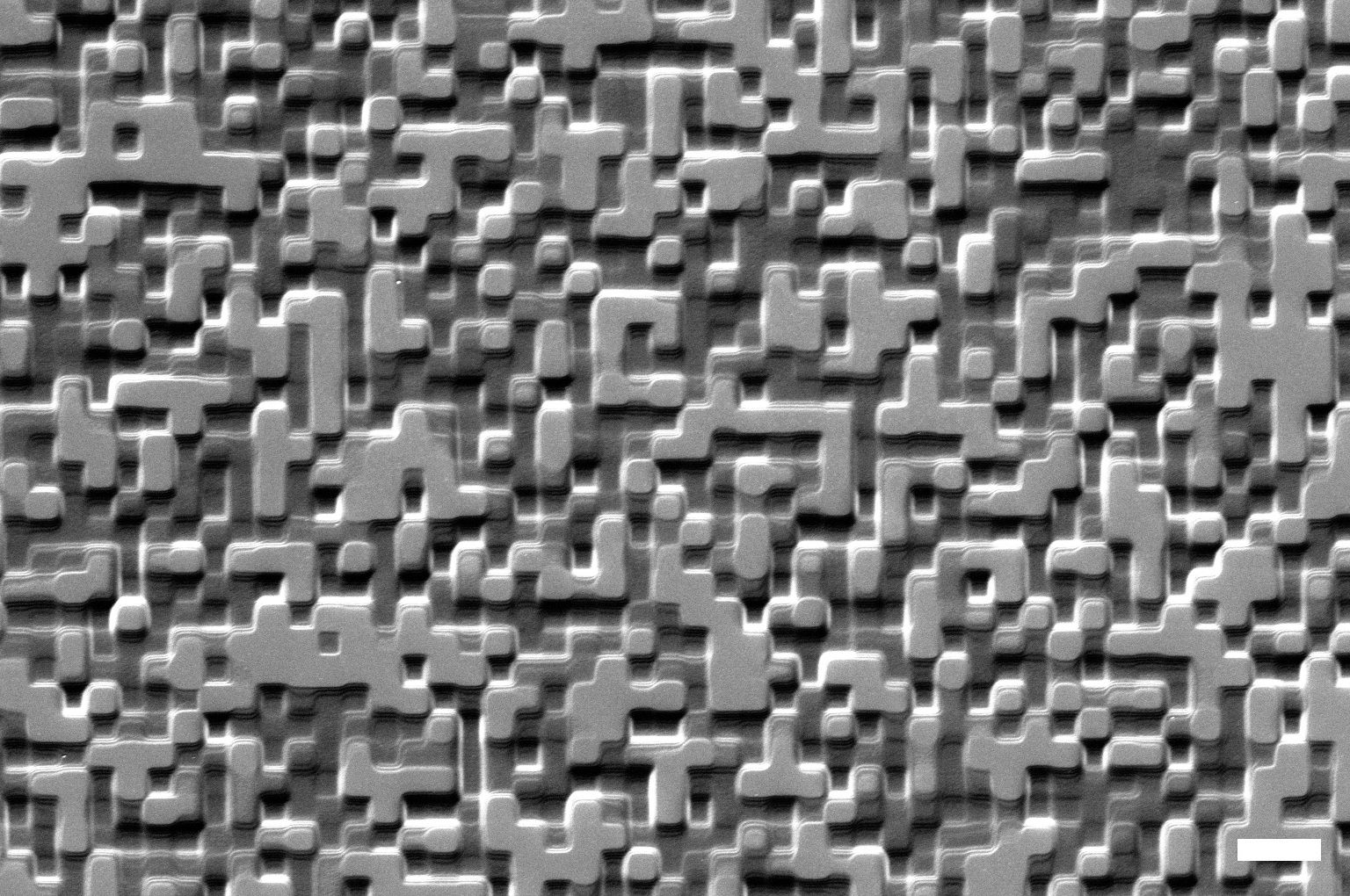
Electron Microscopy and X-Rays
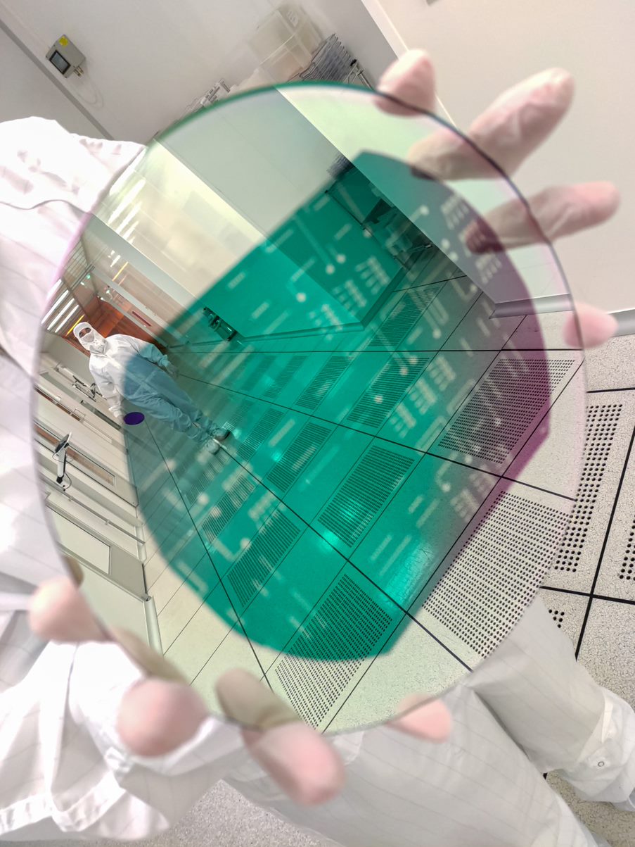
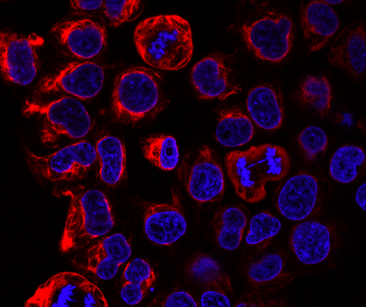
The Advanced Electron Microscopy, Imaging & Spectroscopy (AEMIS) facility is a core multi-user facility that features cutting-edge instrumentation, techniques and expertise required for the characterisation of samples in the physical and life sciences.
This facility focuses on materials and biological research, the development of novel techniques and instrumentation, as well as providing training, technical support and consultation in the areas of electron microscopy and spectroscopy. The centre facility houses several electron microscopes that can probe the physical, electronic, biological, and chemical structure of matter down to the atomic scale.
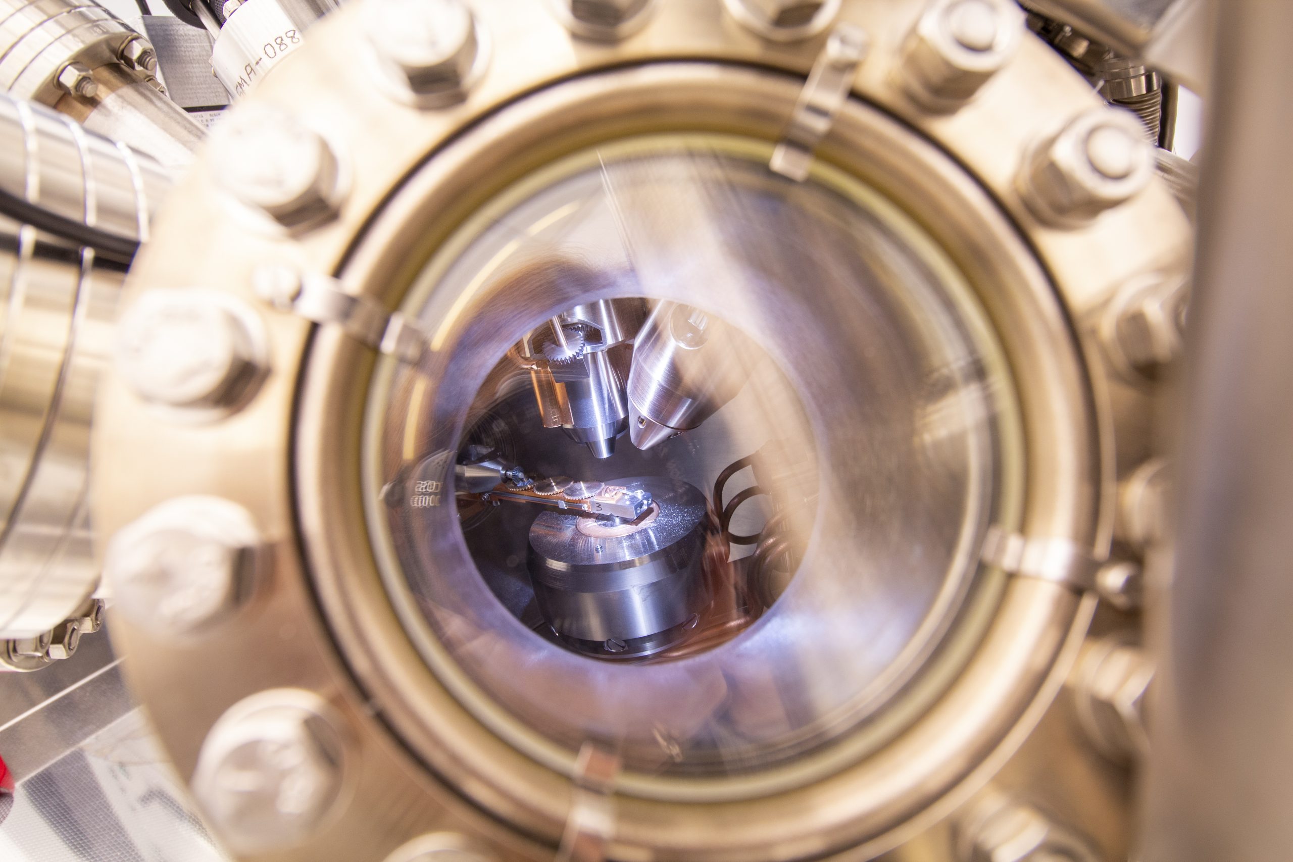
These instruments are coupled with advanced in-situ holders, in which the environment is controlled to match near-realistic conditions of operation, and the sample’s behaviour is recorded dynamically in real time. State-of-the-art support facilities also are available, including standard specimen preparation equipment and an image analysis laboratory.
Probe-Corrected FEI Titan G2 80-200 kV ChemiSTEM
Double-Corrected FEI Titan G3 Cubed Themis 60-300 kV
JEOL JEM 2100 80-200 kVNational Advanced Microscopy Network for Health and Life Sciences – CryoEM-PT
FEI Helios NanoLab 450S DualBeam – FIB with UHREM FEG-SEM
FEI Quanta 650 FEG Environmental SEM (including Peltier and Heating Stage)
Thermo Scientific Escalab 250 Xi
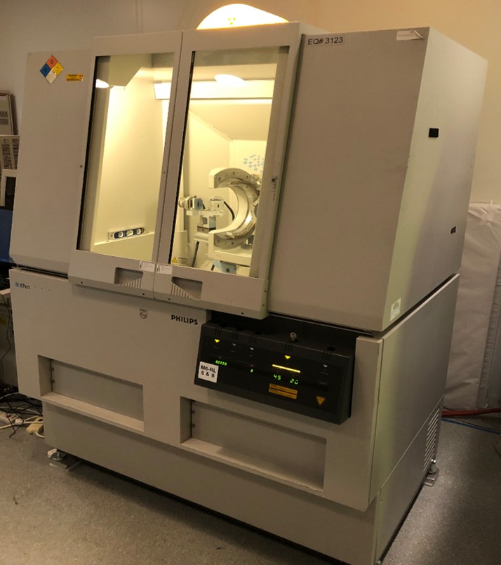
XRD X’Pert PRO Powder or Thin Film X-ray diffraction – an analytical technique used for phase identification and quantification of crystalline materials. It provides information on unit cell structure parameters and crystallographic orientation. Also used for contaminant detection and analysis. Grazing Incidence X-Ray Diffraction can also be done.
Reciprocal Space Mapping – is used to identify crystallographic orientation, composition, stress, and mismatch of heterostructures.
X-ray Reflectivity (XRR) – is used to identify film thickness, density, and interface roughness.
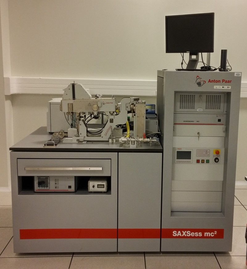
Anton Paar SAXSess mc2 – Measures scattering profiles providing a wide range of information about the structure and properties of the materials Nanoparticle size distribution
Particle shape
Particle structure (e.g. core-shell)
Specific surface area
Agglomeration behaviour of nanoparticles
FEI MEMS-based TEM heating and electrical holder
2 Gatan Cryo-TEM holders (FEI and JEOL microscopes)