Observing Nanovolcanos in the Nanotopologies of Droplets
October 15, 2020

A new method to display the 3D map of nanostructured materials was developed using nanophotonics effects at INL. The new approach is contact-free and therefore does not disturb the object being imaged. The team calls the method – CLeANFIT.
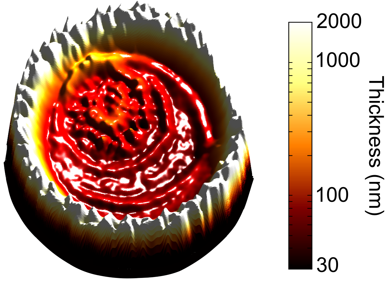
Being contactless is the cherry on top of the cake since it allows mapping the surface nanotopologies even at the liquid interface. When studying an evaporating droplet a nanovulcano landscape was observed.
CLeANFIT is a straightforward tool which may find wide applications in any industry with the need for precise surface or micro/nanostructure characterization.
Imagine, ultrathin nanocoatings need to be formed on a surface to prevent fingerprints or onto an implant to prevent bacteria growth. The relevant manufacturing industry needs highly refined control over the thickness of the ultrathin film deposition, which is an extremely difficult process, particularly at the nanoscale level.
Measurable progress has been done in the fields of materials and coatings to overcome the boundaries between the micro- and nanoscale. However, the characterization of such structures is still restricted to conventional tools, which are known for their macro- and microscale-limited accuracies.
Researchers from the Ultrafast Bio- and Nanophotonics group at INL, could resolve nanometre thicknesses with the help of light-matter interactions. Although many research works tried to overcome such nanoscale challenges with similar interactions, the major obstacle of light diffraction hinders their attempts.
CLeANFIT, provides a novel and promising technique that pushes the boundaries beyond sub-micrometre able to resolve thicknesses down to sub-100 nm scales. To conquer the light diffraction limitations, a particular fluorescence characteristic has been used to identify the distance of the fluorescent matter relative to a nanosensing substrate underneath.
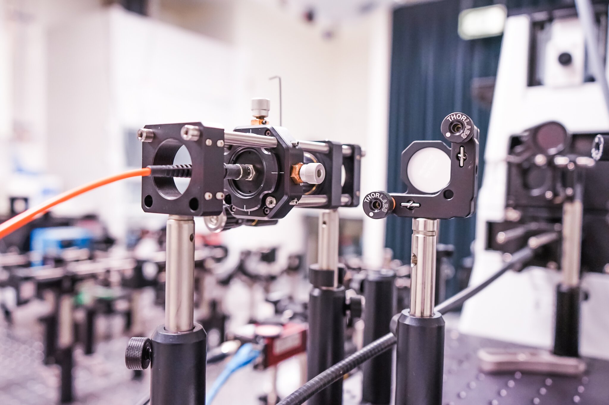
CLeANFIT is composed of three main components: a fluorescence lifetime microscope, fluorescent matter, and a nanosensing substrate. Ricardo Adão, a co-first author of this work, explains that “such setup allied to a physical model of nanophotonic concepts and a numerical algorithm, converts a fluorescence lifetime microscope into a powerful tool for nanometrology applications”.
As a simple demonstration, Ima Ghaeli, the first author of this study, highlights that “the technique can be used to reveal natural fundamental phenomena involved in the evaporation of droplets”. They could not only visualize and quantify the evaporation process while recording the droplet thickness until it dried but also obtain a 3D map of the nanovolcano-shaped patterns that remained on the surface after drying.
The original publication on the nanometrology methodology can be found in the prestigious “Advanced Materials Interfaces” Wiley journal*, and an associated patent application has been filed.
The future plan according to Jana Nieder, who led this study, is to explore the CLeANFIT technique not only using gold but novel substrate materials to explore new sensing ranges and to implement the patent-pending method into industry-standard manufacturing technologies to drive innovation in nanoscale coatings and nanopatterning, such as nanoimprint, inkjet printing, lithography e.g. for electronics, optics, and medical technologies.
Reference
* “CLeANFIT – Contact‐Less Axial Nearfield‐Based Fluorescence Imaging Topography: A Method for 3D Micro‐ and Nanotopography Characterization”, Ima Ghaeli; Ricardo M. R. Adão; Jana B. Nieder, Advanced Materials Interfaces, Pub Date:2020-10-05, DOI: 10.1002/admi.202000581