INL develops novel thermometer to map temperature inside of biological cells
June 7, 2019
A team of researchers from INL – International Iberian Nanotechnology Laboratory developed a new technique that allows measuring the inner temperature of biological cells, opening the door to new applications in the fields of diagnostics and treatment of several diseases, namely cancer. Biological organisms are based on cells – complex machinery optimized to support the specific body functions. Every form of cell activity, like cell division, gene expression, enzyme reaction, metabolism and pathological states are marked by temperature changes.
A balance of biochemical reactions overall leads to a characteristic body temperature allowing optimal function. Everyone knows that checking the body temperature allows helpful feedback to detect inflammation or fever. Diseases often start from a single defect cell and abnormal metabolic activity of such cell may affect its temperature. This is observed for cancer cells, which typically have increased cellular temperatures. This is why cellular temperature techniques may help to identify early-stage diseases. There have been nanothermometers for intracellular applications reported before but mostly requiring very specialized labels or laboratory equipment.

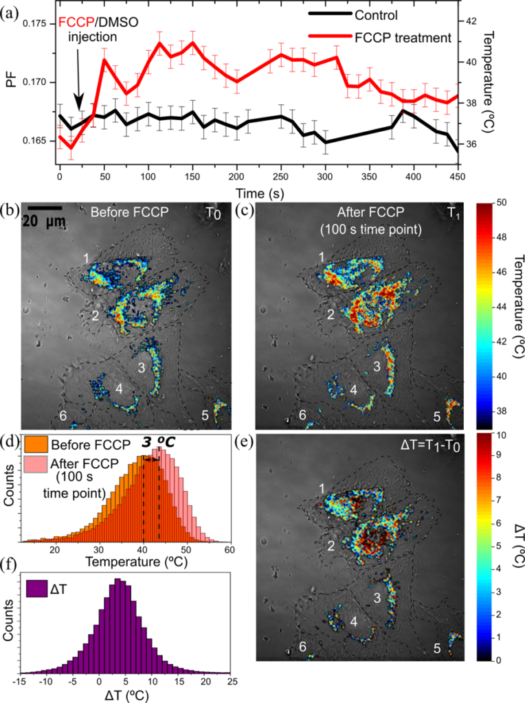
In this study published by an INL research team in the nature group journal “Scientific Reports,” the researchers chose the well-known green fluorescent protein GFP as a source for temperature sensing. The scientists that made the discovery and first deployments of GFP as labelling agent were honoured by a Nobel prize in 2008, as this versatile labelling technique entered rapidly cell and biomedical research laboratories worldwide and let to major discoveries. Commercially several GFP based labelling kits are available for biological or medical analytics. Here for the first time, the INL researchers use the optical properties of GFP to assess the intracellular temperature. The optical parameter used relates to a temperature-dependent spectral shift, to quantify this shift the INL researchers defined a new measure – which was called the “peak fraction parameter”.
According to Oleksandr Savchuk, first author of the study, “The discovery that the peak fraction parameter can be used to measure temperature was performed on a simple solution of the GFP protein. The logical but exciting step was to move this in the context of live cells.”
FROM TEMPERATURE TO THERAPY
Jana Nieder group leading the project, adds that “observing the heat map of a single cell is a fascinating achievement with a rich application potential in fundamental and medical sciences”. We foresee the facile implementation of the technique in standard biomedical labs. As the measurement of the GFP peak fraction parameter can be implemented with simple upgrades on various standard microscopes. The authors furthermore comment: “We hope an uptake of the methodology will lead to future discoveries in cell biology and medical sciences.”
The next envisioned steps are to combine our new intracellular thermometer with the expertise of our nanotechnology experts at INL working on hyperthermia cancer therapies. Hyperthermia treatments aim at locally in raising the temperature to kill specific affected tumour tissues.
On a more fundamental cell biology level we believe the new access to temperature information will enable the observation of disease-specific patterns that may relate to abnormal metabolic activity. The expected new knowledge build-up may trigger new discoveries related to understanding diseases and developing new therapies.
*Original article: https://doi.org/10.1038/s41598-019-44023-7
GFP fluorescence peak fraction analysis based nanothermometer for the assessment of exothermal mitochondria activity in live cells
Oleksandr A. Savchuk, Oscar F. Silvestre, Ricardo M. R. Adão & Jana B. Nieder
Scientific Reports 9, Article number: 7535 (2019)