SyncRGB-FLIM: the new technique to improve biomedical imaging
March 26, 2019
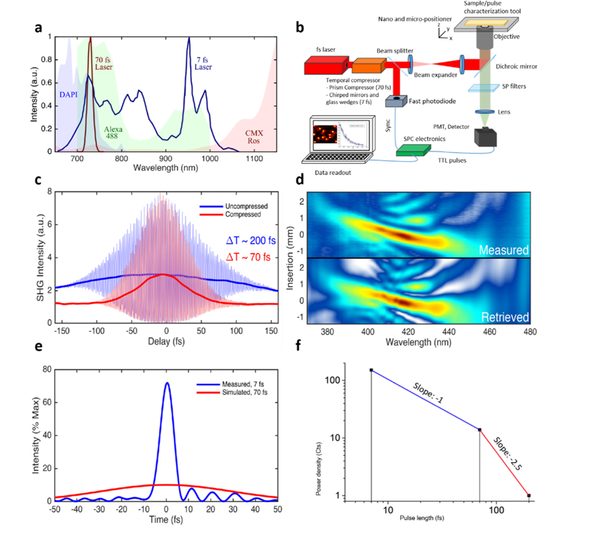
Image © 2019 Optical Society of America under the terms of the and may be used for noncommercial purposes only. Report a copyright concern regarding this image.
A new bio-imaging method for cellular diagnostics named SyncRGB-FLIM was developed in collaboration between INL – International Iberian Nanotechnology Laboratory, the University of Porto and the company Sphere Ultrafast Photonics. SyncRGB-FLIM allows synchronous observation of fluorescent labels with visible emission in the Red, Green and Blue at the same time – this capability is reflected in the name “SyncRGB”. Cellular diagnostics are regularly used to support therapy choices for major health issues faced by the World today, including cancer and neurodegenerative diseases. SyncRGB-FLIM presents an innovative combination of state-of-the-art photonic technologies used to provide an easy-to-use imaging method for fast and advanced clinical and medical imaging. Our research and development aimed to minimise the required technical support of the technique for daily use in hospitals.
The specialised ultrafast laser designed and used for the SyncRGB-FLIM method emits light in very short pulses lasting for only a few femtoseconds (a millionth of a billionth of a second!). According to the famous Heisenberg principle, the shorter the light pulse, the more colours it contains. The ultrafast laser used in this work is so fast that it simultaneously emits coherent light ranging from the infrared to the visible. The colour tuning required by traditional lasers is not necessary here, resulting in faster, more precise and minimally invasive imaging.
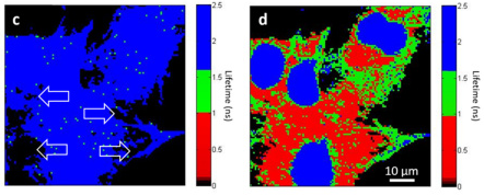
After excitation of the biological samples with this specialised laser source, all labels emit signals at the same time. The signals are all recorded with a single detector, and there is the need to distinguish the origin of the signals. To unmix the signals and distinguish the different RGB dyes, a specific property of the labels is used. This property is called the fluorescence lifetime and requires temporally resolved detection of the signal on a fast time scale of nanoseconds (a billionth of a second). This detection scheme is part of the SyncRGB-FLIM technique, indicated by the letters FLIM standing for Fluorescence Lifetime Imaging Microscopy. The next step for the SyncRGB-FLIM method is to present a full turn-key solution, allowing to taking the system out of the lab and into a real-world medical environment providing a powerful novel technology for cellular and tissue diagnostics to clinicians in hospitals.
The scientific paper about this discovery was just released on Biomedical Optics Express.