
Food Quality and Safety
The main objective of the Food & Quality Research Group is the development of analytical approaches based on the combination of molecular biology (mainly DNA based methodology) and nano and microfabrication technology in order to provide the food industry and control laboratories with reliable analytical tools.
Following this objective, the methodology is based on working on very specific analytical needs and on using a modular approach for each of the steps of the analytical process. This approach, help us to evaluate and to choose the best method in each case, to have a sound integrated final product, and at the same time a wide-range of intermediate products that can be used by themselves to solve specific analytical challenges.
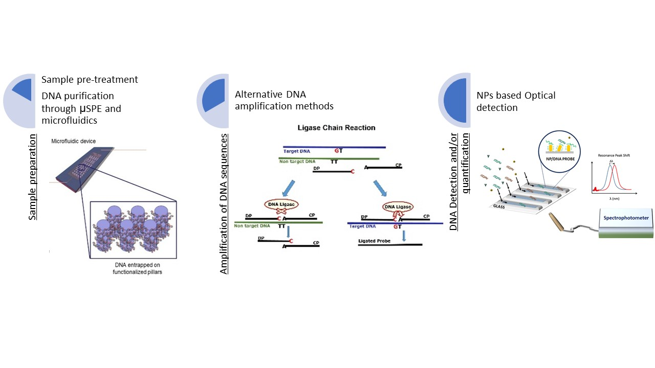
Figure 1 summarizes the overall approach and research lines. Our topics of interest involve the detection of foodborne pathogens, the detection of allergenic ingredients in food products and food authenticity.
Research Lines:
- Sample preparation:
Sample preparation is the series of steps required to transform a sample to a form suitable for analysis, the reliability of the conclusions drawn from food analysis greatly depends upon on this step. We work on: (i) the development of pre-treatment steps in order to overcome some of the limitations associated with food analysis and (ii) on the development of tailored, miniaturized, automatized and faster sample preparation techniques. Microscale solid phase extraction (µSPE) is used for on-chip DNA extraction and purification, being possible to put in contact a higher volume of initial binding material with the solid phase and recover the DNA in a lower volume during the elution phase. This feature allows to concentrate the DNA when minute amounts are present in the sample (e.g. olive oil, wine), for complex matrixes such as processed foodstuff and for environmental samples (e.g. water samples).
- Alternative DNA amplification methods:
Food & Quality Safety Research Group is working on new amplification techniques, in their combination with NPs and on the evaluation of DNA based analytical methods for food analysis. We work on isothermal amplification techniques, such as Loop-Mediated Isothermal Amplification (LAMP), and Recombinase Polymerase Amplification (RPA), specially interesting for miniaturization purposes. Other alternative techniques currently being used include Ligation Chain Reaction (LCR) which allows to distinguish very closely related organisms and high similar DNA sequences.
- Nanoparticle-assisted DNA analysis:
The use of nanomaterials for DNA analysis has the potential of providing increased sensitivity, multiplexing capabilities, and reduced costs. Exploiting the features of nanoparticles (NPs) is considered to be a good alternative to foster the potential of diagnostics and analytical method development. NPs, such as gold NPs (AuNPs) and gold nanorods (AuNRs) are being used for DNA detection taking advantage of their optical properties.